Highlights

  • The dorsal anterior cingulate cortex (dACC) causes motor performance deterioration due to anxiety
  • Transcranial magnetic stimulation of the dACC reduces the performance deterioration
  • This method could potentially be used to help athletes and musicians overcome anxiety
Researchers in the Center for Information and Neural Networks (CiNet), the National Institute of Information and Communications Technology (NICT, President: Hideyuki Tokuda, Ph.D.) and the Centre national de la recherché scientifique (CNRS), France used fMRI to discover a new neural mechanism involving the dorsal anterior cingulate cortex (dACC) to explain how anxiety deteriorates physical performance. Moreover, the performance deterioration was rescued by suppressing brain activity with transcranial magnetic stimulation to the dACC. The findings would provide a new therapeutic strategy for athletes, musicians and other performers susceptible to anxiety during performance. The paper was published online in Nature Communications on September 19, 2019.
 

Background

Athletes, musicians and other performers, be they professionals or amateurs, are required to conduct rapid complex movements that can be affected by anxiety. Influential self-focus theory postulates that the motor skills required for these actions become automatic and unconscious during learning, but anxiety causes an interference between conscious and unconscious processing that can negatively impact the performance. However, no behavioral or brain data has confirmed the theory. Such data would provide therapeutic targets that negate the effects of anxiety on motor performances.

Achievements

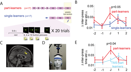
Fig. 1 Reduced performance under anxiety correlated with activity in the dACC.
[Click picture to enlarge]
Principle Investigator Masahiko Haruno (NICT), with Dr. Gowrishankar Ganesh (CNRS) and Dr. Takehiro Minamoto (Shimane University) conducted fMRI by designing a novel behavioral task and found a correlation between activity in the dACC and the motor performance deterioration due to anxiety. The application of TMS to suppress dACC activity rescued the deterioration, providing the first direct evidence that suppressing anxiety-stimulated regions could reduce performance deterioration.  
In general, specific motor skills, such as those used in tennis or playing the piano, require repeated practice to memorize the motions and their order. In the new study, the researchers simulated this practice on a computerized 10-step rapid button press task. Part-learners learned the task in two parts, one 6 steps long and the other 4 steps long. Finally, they practiced all 10 step at once. Single-learners, on the other hand, learned the 10-step rapid button presses together without breaking them into parts. After the learning, both groups of learners were asked to conduct the entire 10-step task and given an electrical shock if they made a mistake during the performance (Fig. 1A, anxiety test session). 
Part-learners proved more adept at learning the task in the training sessions based on the speed and number of errors with which they completed the task. However, when the anxiety test session started, their performance dropped noticeably to a level worse than single-learners (Fig. 1B, J represents the junction of the two parts). This finding is consistent with the self-focus theory, which would expect performance to decline with anxiety. 
fMRI revealed that the part-learners showed increased activity of the dACC at the time of the junction in the test session (Fig. 1C). Applying TMS to this region for 5 minutes (1 Hz) prior to the test session (Fig. 1D) eliminated the performance degradation in part-learners (Fig. 1E). On the other hand, part-learners who received sham TMS did not show improvement (see Supplementary for details).
These experiments show for the first time the brain region responsible for performance degradation caused by anxiety and a possible therapeutic strategy using TMS. 

Future research

In addition to more study of the dACC neural circuits that connect anxiety to performance degradation, the group will investigate how TMS can enhance the performance of athletes, musicians and other performers.

Information of the article

Journal: Nature Communications
DOI: 10.1038/s41467-019-12205-6
Title: Activity in the dorsal ACC causes deterioration of sequential motor performance due to anxiety
Authors: Gowrishankar Ganesh, Takehiro Minamoto, Masahiko Haruno
 
*Gowrishankar Ganesh is a senior researcher at the Centre national de la recherché scientifique (CNRS), France. 

Project funding

This research was funded in part by JST-CREST for the “Prediction of social attitudes and stresses based on social neuroscience and natural language processing” to Masahiko Haruno under the “Intelligent information processing systems creating co-experience knowledge and wisdom with human-machine harmonious collaboration” project and JSPS KAKENHI grant for “Brain information dynamics underlying multi-area interconnectivity and parallel processing”.

Supplementary

Details of the experimental findings

In this study, researchers conducted three types of experiments (behavioral, fMRI, and TMS) to examine performance degradation under anxiety and underlying brain activity, thus finding a causal relationship with the dACC.
 

[Experiment 1: behavioral]

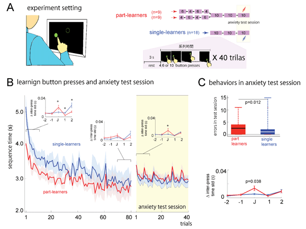
Fig. 5. Design and results of experiment 1 (behavioral).
Several skills like those performed by tennis players and pianists are first learned in parts and then assembled together. A task to model this process was applied to 18 subjects, who were asked to touch a 10-button panel in accordance to the presentation of 10 green circles as fast as possible without making errors. The subjects were broken into two groups: part-learners and single-learners. The part-learners learned the 10-step sequence in sets of 6 steps and 4 steps and then as one set of 10 continuous steps. The single-learners learned the entire sequence as one set (Fig. 5A). Next, the subjects participated in a test session of the learned sequence, but under the threat of a weak electric shock for an error or taking too long to press the button. Too long a time was defined as 1.5 times the average time for the last 10 practice trials and unknown to the participant. The intensity of the shock was adjusted for each subject. The order of the part- and single- learning was randomized across subjects.
Fig. 5B shows the length of time to conduct the sequence for both groups during the practice (80 trials) and test (40 trials) sessions. While both got faster with more practice sessions, part-learners remained consistently faster. When the test session was conducted with the electric shock, however, part-learners actually took longer than single-learners. 
Next, the subjects were observed for the time between button presses during a sequence. This time became smaller as the subject learned the sequence in the practice session. However, the addition of the electric shock in the test session caused an obvious change (Fig. 5B and C). The letter J in the figures indicates the break of the two sub-sequences learned by the part-learners. The numbers 1 and 2 indicate when the first and second buttons were pressed in the second sub-sequence, and the numbers -1 and -2 indicate the last and penultimate buttons pressed in the first sub-sequence. Early in the practice session, the part-learners showed high variability, but that gradually disappeared with more trials (Fig. 5B insets, red lines; single learners showed no difference because the sequence was learned as one continuous set), suggesting the task had become automated. However, during the test session, this variability returned (Fig. 5C). This observation is consistent with the self-focus theory, in which the automated motor actions are disturbed by conscious interference. Overall, part-learners showed more errors than single-learners and more variation at the J junction. 
From these experiments, it could be concluded that the effect of anxiety on performance depends on the method of learning the performance, that the effect is consistent with the self-focus theory, and that the effect of anxiety was strongest at the junction.
 

[Experiment 2: fMRI]

fMRI experiments were then conducted to identify the brain regions associated with the degradation seen in experiment 1. 
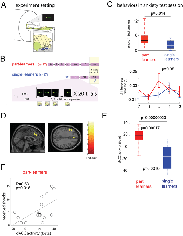
17 new part-learners and 17 new single-learners were employed, and the design of experimental 1 was modified to accommodate the use of the fMRI machine. In this case, only 20 sessions were done, and the user only needed to press one of three buttons using an optical key pad (Fig. 6A and B).
 
Fig. 6. Design and results of experiment 2 (fMRI).
Consistent with experiment 1, part-learners made more errors in experiment 2 during the test session and showed greater variation at junction J (Fig. 6C).
fMRI indicated that the activity of the dACC correlated with the change seen at junction J, which was greater in part-learners than in single-learners (Fig. 6D and E). Furthermore, there was a significant correlation between the activity of the dACC and the number of electrical shocks received by the part-learners (Fig. 6F). Thus, it was suggested that the performance decline of part-learners was attributed to the activity of the dACC.
 

[Experiment 3: TMS]

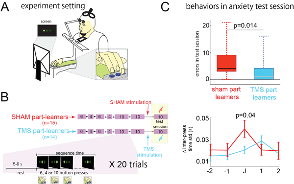
In experiment 3, TMS was performed on the dACC only for newly recruited part-learners (Fig. 7A and B). The condition was the same as experiment 2 except with the TMS (1 Hz, 320 pulses, 1.2 RMT) performed just before the test session. In contrast with TMS part-learners, SHAM part-learners had a plate placed between the TMS double-cone coil and head so that the dACC was not stimulated. 
TMS part-leaners who underwent TMS showed fewer errors than SHAM part-learners (Fig. 7C). In fact, the number of errors was comparable with single-learners. Additionally, they showed no variation increase at junction J (Fig. 7C). These results confirm a causal relationship between dACC activity and performance degradation. 
Overall, this study designed a novel behavioral task to investigate how anxiety compromises rapid sequential motor performance. fMRI attributed this effect to increased activity of the dACC. Finally, TMS to the dACC prior to the anxiety could recover the performance. These results show a causal relationship between dACC activity and performance degradation.
 
Fig. 7. Design and results of experiment 3 (TMS).
Appendix
All subjects were informed of the details of the experiments and gave their consent prior to participating. The experiments were approved by an ethics committee at NICT.

Glossary

fMRI(functional Magnetic Resonance Imaging)
A derivative of magnetic resonance imaging (MRI), fMRI is a non-invasive imaging technique that measures magnetic resonance to correlate cerebral blood flow and brain activity.
Dorsal anterior cingulate cortex (dACC)
A region of the brain whose activity is associated with cognitive control, social behavior and resolution of conflicts. It is responsible for regulating several cognitive and emotional functions and has neural connections with the amygdala, and other regions of the prefrontal cortices. In the current study, the dACC was found to activate when learned cognitive skills were connected consciously under a state of anxiety (self-focus theory). The results suggest that cognitive control, which is normally beneficial to motor performance, can also be detrimental. 
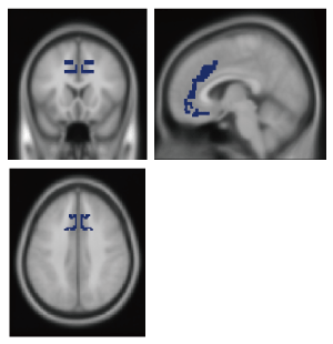
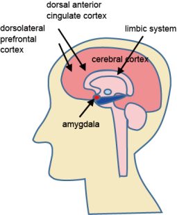
Fig. 2 shows the location of the dACC (Brodmann area 32), and Fig. 3 shows an illustration of its relative position in the brain.
 
Fig. 2. Location of the dACC.
Fig. 3. Illustration of the brain and relative position of the dACC.
Transcranial Magnetic Stimulation (TMS)
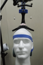
Fig. 4. TMS double-cone coil.
TMS is a non-invasive method that uses a dynamic magnetic field to generate a weak current that activates or suppresses targeted neurons. It is commonly used to investigate neural networks. 
Past research has shown that repetitive TMS (rTMS) at 1 Hz reduces neural activity and can have a therapeutic effect on patients with Parkinson’s disease, dystonia, or depression. 
In this study, a double-cone magnetic coil was used for the TMS (Fig. 4), which could suppress cells as far as 3 cm from the head surface. 
In 2009, the International Federation of Clinical Neurophysiology released guidelines on safe TMS delivery, and in 2011, the Japanese Society of Clinical Neurophysiology translated these guidelines to Japanese. The guidelines state that up to 15,000 pulses can be applied in one week at a maximum frequency of 10 Hz and stimulation intensity 1.2 times the resting motor threshold (RMT).
In the current study, the TMS conditions were 1 Hz, 1.2 RMT, and 320 pulses applied one time.

Technical Contact

Masahiko Haruno
Neural Information Engineering Laboratory
Center for Information and Neural Networks
NICT

Tel: +81-80-9098-3239

E-mail: mharuno_atmark_nict.go.jp

Media Contact

Sachiko Hirota
Press Office
Public Relations Department
NICT

Tel: +81-42-327-6923

E-mail: publicity_atmark_nict.go.jp